
Figure 2.
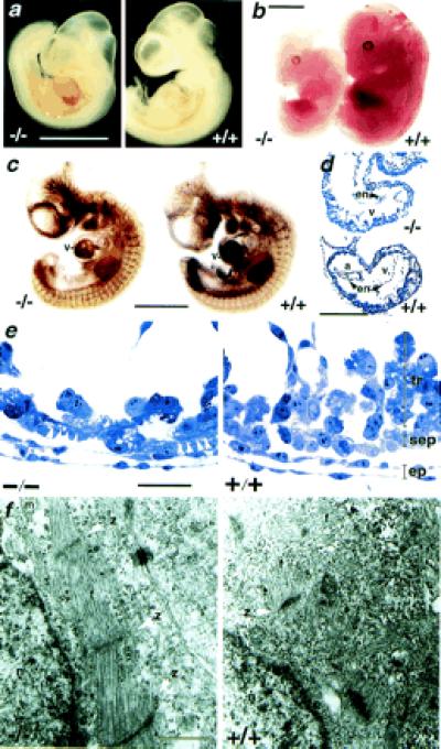
Morphology of 5-HT2B mutant embryos. (a) 5-HT2B-deficient embryos at 10.5 dpc exhibit a typical bleeding into the pericardial cavity. (b) At 12.5 dpc, the mutant embryos are smaller and paler than their wild-type littermates. (c) Whole-mount immunohistochemical staining of 10.5 dpc embryos for developing blood vessels with platelet endothelial cell adhesion molecule antibody revealed no gross defects in vascular pattering compared with age-matched wild-type littermates but reduced staining in heart ventricle (v). (d) Semithin sagittal sections of 9.5 dpc embryos demonstrate a severe reduction in the thickness of the ventricle (v) including both the compact zone and the trabeculae in 5-HT2B-deficient embryos. a, Atrium; en, endocardium. (e) Higher magnification shows a reduction of trabecular cells (tr) in the mutant heart, whereas elongated cells (white arrowhead) are visible in the compact subepicardial layer (sep). Epicardial cells (ep) are normally developed. (f) TEM analysis of these embryos reveals the abnormal sarcomeric differentiation within the subepicardial layer in all observed mutant heart (n = 6) but not in wild-type heart. z, Z band of the sarcomeres; f, actin fibers; m, mitochondria; and n, nucleus. Genotype designations are +/+, wild type; and −/−, homozygous mutant. (Bars for a–c = 500 μm; d = 100 μm; e = 5 μm, and f = 0.5 μm.)