
Figure 4.
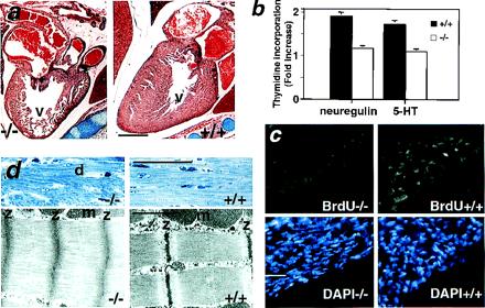
Heart defects in postnatal 5-HT2B mutant mice. (a) Histological analysis of newborn mutant hearts shows an obvious loss of ventricular (v) mass (hypoplasia). (b) Thymidine incorporation of isolated cardiomyocytes from newborn heart in response to 5-HT and neuregulin (heregulin) is significantly reduced in the 5-HT2B mutant, indicating that myocyte proliferation is impaired and this effect is cell autonomous. The thymidine incorporation rate is expressed as fold increase over basal ± SEM (−/−, n = 3; and +/+, n = 3). (c) In vivo, BrdUrd incorporation (green) is almost undetectable in the heart of 5-HT2B newborn mutant (Left), whereas BrdUrd incorporation was observed in the compact zone of control newborn heart (Right). Staining with 4′,6-diamidino-2-phenylindole of the same sections localizes the cell nuclei (blue). (d Upper) Semithin sections of heart in 6-week-old mutant shows abnormal morphology, including degenerating fiber (d) (Left). (d Lower) TEM of ultrathin sections shows that, in mutant heart, the sarcomere lengths are greatly reduced, the M-lines and the I-bands are indistinguishable, and the A-bands occupy the entire length between Z-bands (z) which themselves are thickened (Left) compared with wild-type age-mated heart (Right). m, Mitochondria. (Bars for a = 100 μm; c and d Upper = 20 μm; and d Lower = 0.5 μm.)