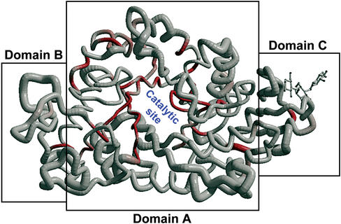
Figure 2.
Cα trace of the structure of AMY1 coloured from white to red according to sequence similarities (from low to high). The radius of the Cα tube is proportional to rms deviation after superimposition of the structure of AMY1 with its homologues. The most conserved region is the beta-barrel of domain A containing the catalytic site. The thio-maltodextrine molecule bound to the domain C is shown in ball-and-sticks. Figure prepared with BOBSCRIPT (12).

