
Figure 4.
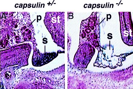
Histologic analysis of the developing spleen at E13.5. A shows a section through the developing spleen and surrounding tissues of a capsulin+/− embryo at E13.5. LacZ staining is apparent in the spleen. B shows a section through a capsulin−/− embryo at the same level as in A. Note the region of the developing spleen is virtually devoid of cells, whereas the adjacent pancreas and stomach appear normal. p, pancreas; s, spleen, st, stomach.