
Figure 2.
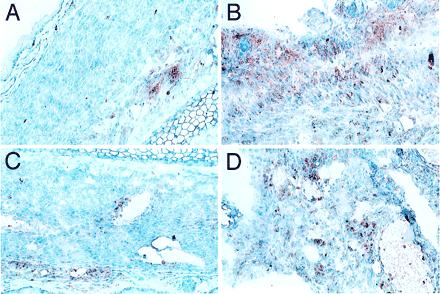
Immunohistochemical analysis of mH-mismatched neonatal heart grafts transplanted in HSC chimeric mice. A BALB/c mouse engrafted with BA HSCs was killed on day +82 and day +51 after transplantation of heart grafts from a DBA.2 (A and C) and a C3H.SW (B and D) neonate into the pinna of contralateral ears, respectively. (A and B) Staining for macrophages and monocytes (α-Mac-1). (C and D) Staining for T cells (α-CD3). Note healthy appearance of myocardial tissue and mild peripheral involvement with inflammatory cells in the DBA.2 graft as compared with the C3H.SW, which has heavy mononuclear cell involvement and evidence of dying myocardial cells.