
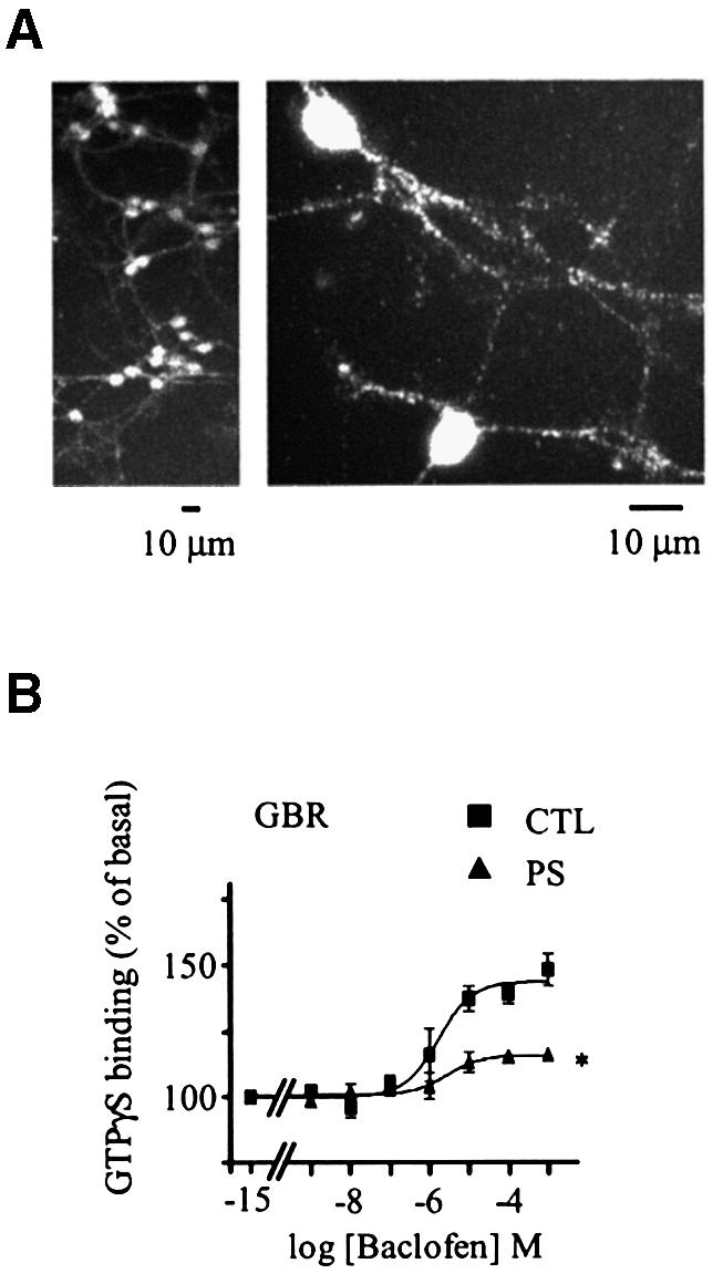
Fig. 3. Desensitization of GBR in cerebellar granule cell. (A) Endogenously expressed GBRs were detected in cerebellar granule cells (GCG) by immunofluorescence microscopy. Permeabilized cells were immunolabelled using a polyclonal antibody raised against native GBR2. (B) Baclofen-stimulated [35S]GTPγS binding was measured in membranes derived from CGC, pre-stimulated (PS; triangles) or not (CTL; squares) for 60 min with the selective agonist (1 mM baclofen). Data represent the mean ± SEM of three independent experiments performed in triplicate. *P < 0.05.