
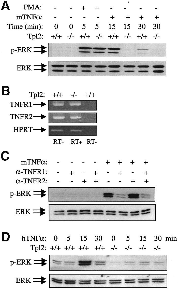
Fig. 4. ERK activation by TNF-α in macrophages depends on signals transduced via Tpl2. (A) Representative western blots of Tpl2+/+ and Tpl2–/– bone marrow-derived macrophage (BMDM) cell lysates, harvested at the indicated time points before and after PMA or murine TNF-α (mTNFα) stimulation, was probed with antibodies to phosphorylated ERK (upper) or total ERK (lower). (B) Tpl2+/+ and Tpl2–/– BMDMs express both TNFR1 and TNFR2, as determined by RT–PCR. Amplification of the housekeeping gene HPRT serves as a cDNA synthesis and amplification control, and the lack of DNA contamination is confirmed by the absence of amplification in the RT– sample. (C) TNFR1 transduces ERK signaling in mTNF-α-treated mouse BMDMs. BMDMs from Tpl2+/+ mice were pretreated for 30 min with 2 µg/ml blocking TNFR1, TNFR2 mAbs or a combination of these reagents and then stimulated for 15 min with 50 ng/ml mTNF-α. Lysates were examined for phosphorylated or total ERK. (D) The effects of human TNF-α (hTNFα), which exclusively stimulates the mTNFR1, on ERK activation in Tpl2+/+ and Tpl2–/– BMDMs.