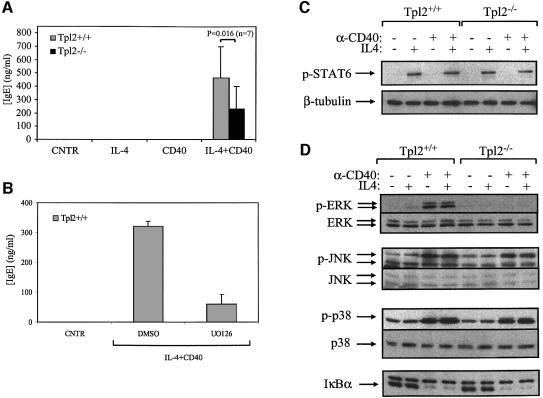
Fig. 7. B cells from Tpl2–/– mice are partially impaired in IgE production following CD40 and IL-4 stimulation. (A) Secreted IgE levels (means ± SD from seven independent experimental B-cell cultures) are reduced in Tpl2–/– B cells stimulated with anti-CD40 plus IL-4. This difference is statistically significant (p = 0.016). (B) Secreted IgE levels (means ± SD from four independent experimental B-cell cultures) are reduced in Tpl2+/+ B cells treated with IL-4 and anti-CD40 in the presence of the MEK inhibitor UO126. The levels of IgE from vehicle control (DMSO)-treated cultures are also shown. (C) CD40 does not affect the levels of STAT6 phosphorylation induced by IL-4. B cells from Tpl2+/+ and Tpl2–/– mice were stimulated with anti-CD40, IL-4 or anti-CD40 plus IL-4 for 15 min, and cell lysates were analyzed for the phosphorylation status of STAT6 by immunoblot analysis (upper). Equal loading was confirmed by probing the same blot for β-tubulin (lower). (D) IL-4 does not modify the MAPKs and NF-κB activation by CD40. B cells from Tpl2+/+ and Tpl2–/– mice were stimulated with anti-CD40, IL-4 or anti-CD40 plus IL-4 for 15 min, and cell lysates were analyzed for MAPK activation using antibodies that recognize either the phosphorylated or total forms of ERK, JNK and p38. IκBα degradation was assessed by immunoblot using an anti-IκBα polyclonal antibody. The experiments in (C) and (B) were carried out on the same cell lysates. This confirmed that both the anti-CD40 antibody and the IL-4 were functionally active when used alone.

An official website of the United States government
Here's how you know
Official websites use .gov
A
.gov website belongs to an official
government organization in the United States.
Secure .gov websites use HTTPS
A lock (
) or https:// means you've safely
connected to the .gov website. Share sensitive
information only on official, secure websites.
