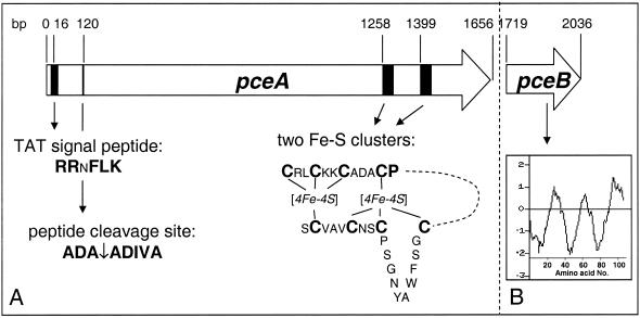
FIG. 6.
Physical representation of the PceA and putative PceB RDases of D. restrictus. (A) PceA and its features, including the TAT signal peptide with a conserved RRxFLK signature, the peptide cleavage site, and two [4Fe-4S] clusters towards the C-terminal end. (B) PceB putative protein and a hydrophobicity plot indicating the presence of three transmembrane α-helices. The Kyle-Doolittle hydrophobicity plot was obtained by using the software Protein Hydrophilicity/Hydrophobicity Search (Bioinformatics Unit, Weizmann Institute of Science, Rehovot, Israel).

