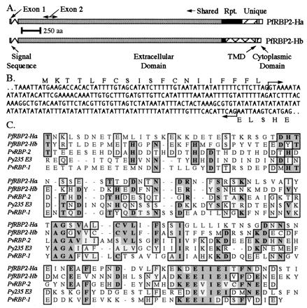
Figure 3.

Structure of the PfRBP2-H genes and their relationship with each other and with other Plasmodium proteins. (A) Schematic of the PfRBP2-H genes, highlighting structural features as marked. Shared regions (gray), repeats (black), and unique regions (hatched) are also noted. (B) Sequence of the 5′ end of the PfRBP2-H genes, showing the boundaries of the intron (arrows) and sequence of the short exon 1 that encodes a signal peptide. (C) clustal alignment of the C terminus of the PfRBP2-H proteins, the P. vivax reticulocyte-binding proteins, and the E-3 member of the P. yoelii p235 family. Identical residues are highlighted in gray and similar residues in light gray.
