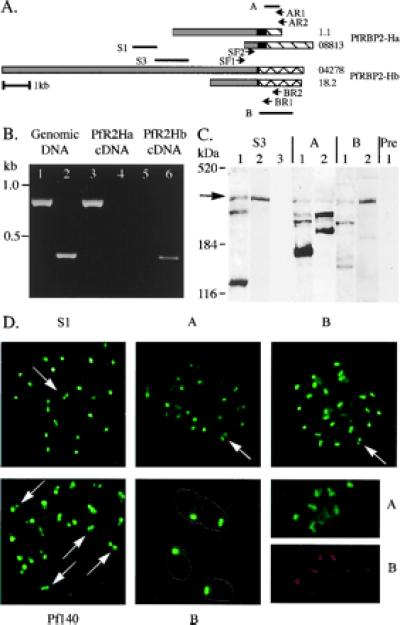
Figure 5.

PfRBP2-Ha and -Hb are expressed at the apical end of merozoites. (A) PfRBP2-Ha (1.1 and 08813) and -Hb (18.2 and 04278) schematic. Gray, black, and hatched boxes define shared, repeated, and unique regions, respectively. Primers used in reverse transcription–PCR are marked (SF1, SF2, AR1, AR2, BR1, and BR2), as are the fragments of the genes expressed to raise antisera (S1, S3, A, and B). (B) Nested PCR was performed by using primers specific to PfRBP2-Ha (SF1/AR2 followed by SF2/AR1, lanes 1, 3, and 5) or -Hb (SF1/BR2 followed by SF2/BR1, lanes 2, 4, and 6), with either FVO DNA (lanes 1 and 2) or cDNA as templates. The cDNA was generated from schizont stage total RNA and primers specific to PfRBP2-Ha (lanes 3 and 4) or PfRBP2-Hb (lanes 5 and 6) for reverse transcription reactions. (C) Western immunoblots prepared with anti-S3 (S3), anti-PfRBP2-Ha (A), anti-PfRBP2-Hb (B), or preimmune (Pre) antiserum as detection agent. Lanes contain either P. falciparum mature schizont SDS extracts (1 and 2) or uninfected erythrocyte ghost preparations (3). Protein size standards from GIBCO/BRL and Coomassie-stained apolipoprotein B (520 kDa) are noted. (D) Immunofluorescence assays performed with antisera against fragments of the shared domain (S1), fragments of the unique regions, anti-PfRBP2-Ha (A) and anti-PfRBP2-Hb (B), and the rhoptry antigen Pf-140, on air-dried, acetone-fixed, P. falciparum-infected erythrocytes. The lower center panel (B) is at a higher magnification (×3,750; others are ×1,250), with the outline of the merozoites marked. The lower right shows costaining of single merozoites with rabbit anti-A serum (A) and rat anti-B serum (B).
