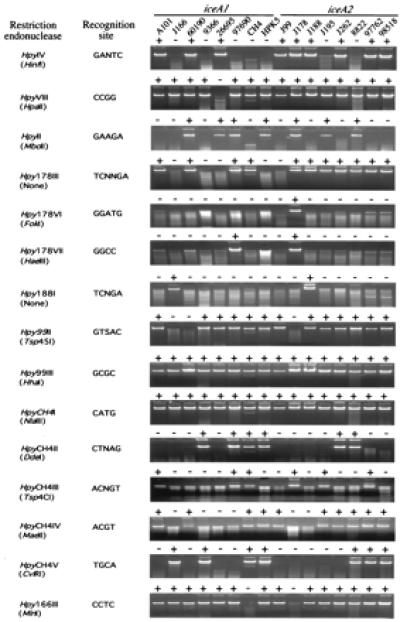
Figure 1.

Digestion of chromosomal DNA from 16 H. pylori strains by 15 different type II REs. The digests, resolved in a 1% agarose gel, are listed in the right panel. “+” indicates digestible by the RE whereas “−” indicates resistant to digestion. The name, recognition site, and prototype of each RE are listed in the left panel. The sources of the chromosomal DNA are listed at the top of the figure. The iceA genotypes of the DNA are also indicated.
