
Figure 2.
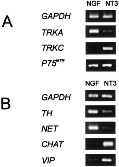
Differential gene expression of sympathetic explants. E12 explants were cultured for 4 days, and relative gene expression levels were determined by reverse transcription-coupled PCR. Amplification of GAPDH was used to normalize the cDNA content of the samples. Photographs are representative examples. Ratios of >20 indicate very strong differences, which were not resolved. (A) Neurotrophin receptor genes. Primer sets used for the amplification of TRKA and TRKC correspond to sequences from the intracellular domains encoding the full-length tyrosine kinases (24, 25). Consistently strong differences between NGF-treated and NT-3-treated cultures were observed for TRKA and TRKC (gene expression ratios 5 ± 2:1, 1:>20). The neurotrophin receptor P75NTR gene was expressed at similar levels (1:1). (B) Neurotransmitter marker genes. Transcript levels for the noradrenergic markers NET (>20:1) and TH (5 ± 2:1) were higher in NGF-treated explants compared with explants with NT3. In contrast, transcripts of the cholinergic marker genes CHAT (1:>20) and VIP (1:15 ± 3) were enriched in NT3-treated compared with NGF-treated sympathetic neurons.