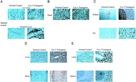
Figure 2.

Immunohistochemical analysis of the tissue distribution of caveolin-3 in normal control mice and caveolin-3 (Cav-3) transgenic mice. Paraffin-embedded tissue sections were prepared and immunostained with anti-caveolin-3 IgG. After extensive washing, bound primary antibodies were visualized by using a peroxidase-based detection method (see Materials and Methods). Note that in normal control mice, caveolin-3 expression is essentially confined to striated muscle tissues [skeletal muscle (A) and heart (B)]. In contrast, in caveolin-3 transgenic mice, caveolin-3 is widely expressed in a variety of different tissues: kidney, fat (C); liver, brain (D); lung, spleen (E). In skeletal muscle (A) and in the heart (B), note that caveolin-3 transgenic mice exhibit overexpression of caveolin-3 when compared with the endogenous expression observed in normal control mice.
