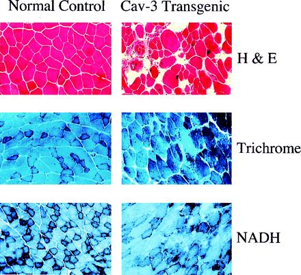
Figure 4.

Histological and histochemical analysis of skeletal muscle fibers from caveolin-3 (Cav-3) transgenic mice. Muscle tissue sections from normal control mice (Left) and caveolin-3 transgenic mice (Right) were stained with hematoxylin and eosin (H&E) (Top), modified Gomori's trichrome (Middle), or NADH-tetrazolium reductase (Bottom). Note that muscle tissue sections from caveolin-3 transgenic mice exhibit dramatic variability in the size of muscle fibers, an elevated number of necrotic fibers and regenerating muscle fibers (i.e., fibers with centrally located nuclei), and significant proliferation of connective tissue.
