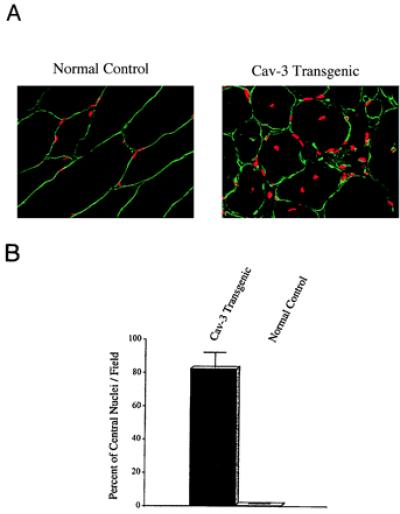
Figure 5.

Caveolin-3 (Cav-3) transgenic mice exhibit an elevated number of skeletal muscle fibers with centrally located nuclei. (A) Immunocytochemistry. Skeletal muscle tissue sections from normal control mice (Left) and caveolin-3 transgenic mice (Right) were immunostained with anti-caveolin-3 IgG to reveal the muscle fiber sarcolemma (plasma membrane) and counterstained with propidium iodide to visualize the nuclei. The resulting merged images are shown to better allow visualization of the position of the nuclei with respect to the plasma membrane. (B) Quantitation of central nucleation. The number of muscle fibers with central nuclei is represented as percentage of the total muscle fibers analyzed. Note that in caveolin-3 transgenic mice ≈82% of the muscle fibers contain central nuclei, whereas ≈0.5% of muscle fibers from normal control mice exhibit central nuclei. These values were determined by examining the morphology of muscle fibers from > 30 different fields.
