
Figure 2.
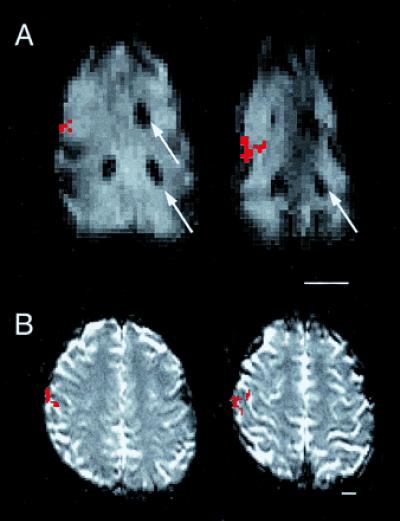
Activity generated in anterior parietal cortex of the anesthetized macaque monkey (A) and the awake human (B) using the same stimulus to stimulate cutaneous receptors on the entire glabrous hand of each species. In both A and B, serial axial EPI MR images are displayed, with the most inferior section on the left and rostral to the top. Active voxels are indicated in red. (A) The activity generated from the applied stimulus was easily related to the gadolinium filled probes (arrows) placed before the commencement of fMRI mapping experiments (see Materials and Methods). The relative location of activation with respect to major sulcal landmarks and activations generated from face stimulation (not shown) is the same in both the anesthetized monkey and the awake human. (Scale bars equal 1.5 cm.)