
Figure 1.
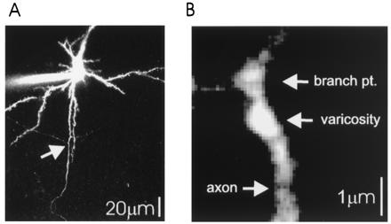
Imaging of axonal arbors of layer 2/3 pyramidal cells. (A) Low magnification image of a neuron filled with 100 μM OGB1, showing the apical dendrite (Top), several spiny basal dendrites, and the primary axon with two primary to secondary branch points (arrow). Note the low fluorescence intensity of secondary axonal branches compared with basal dendrites, consistent with the small diameters of axons. (B) High magnification image of a branch point between secondary and tertiary branches (same axon as in 2C). An axonal swelling is clearly recognizable by large resting fluorescence. Axonal branch points also show relatively large fluorescence.