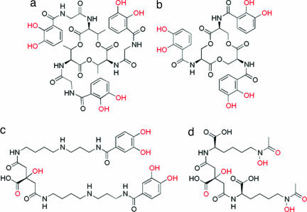
Fig. 1.
Molecular structures of B. anthracis siderophores (a and b) and the corresponding siderophores from Gram-negative enteric bacteria (c and d). (a) BB. (b) PB. (c) Ent.; (d) Aerobactin. The iron-coordinating oxygen atoms (when deprotonated) are indicated in red. All completely sequester the FeIII by six-coordinate binding.

