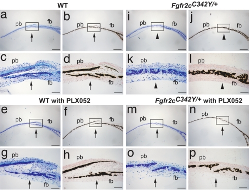
Fig. 5.
Suture fusion in calvaria tissue explants is prevented by treatment with FGFR inhibitor. Calvaria harvested from E18.5-day-old embryos of WT (a–h) and Fgfr2cC342Y/+ mutant (i–p) mice were cultured either with vehicle (0.2% DMSO) alone (a–d and i–l) or with 1 μM PLX052 (e–h and m–p) for 2 weeks. Histological sections of cultured calvaria were made perpendicular to the coronal suture, which passes through the frontal bone (fb) and the parietal bone (pb). Sections were stained with toluidine blue (a, c, e, g, i, k, m, and o), and adjacent sections were stained by von Kossa followed by methyl green counter staining (b, d, f, h, j, l, n, and p). Lower [c, d, g, h, k, l, o, and p (Scale bars: 100 μm)] depicts the higher magnification of the coronal sutures shown in the boxed regions of Upper [a, b, e, f, i, j, m, and n (Scale bars: 500 μm)], respectively. Open (arrow) and fused coronal sutures (arrowhead) are indicated. Fused sutures are observed in untreated calvaria from mutant mice (i–l), and open sutures are observed in calvaria from mutant mice treated with PLX052 (m–p).

