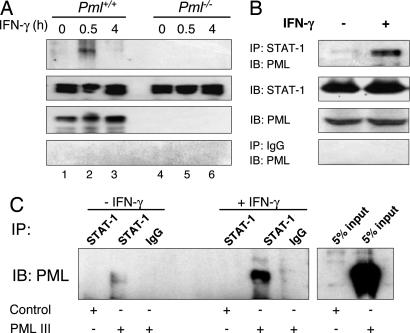
Fig. 4.
PML interacts with STAT-1 in vivo. (A) Immortalized Pml+/+ and Pml−/− MEF were incubated in the absence or presence of IFN-γ for 0.5 and 4 h, and whole-cell lysates were prepared. Cell lysates (1.2 mg) were immunoprecipitated with anti-STAT-1 antibody and subjected to SDS/PAGE for immunoblot analysis with anti-PML antibody. Input samples (5%) were assayed for STAT-1 and PML protein expression by immunoblotting. (B) RAW264.7 cells were either untreated or treated with IFN-γ for 0.5 h and processed as described in A. (C) Pml−/− MEF transfected with control vector or the PML III expression vector were either untreated or treated with IFN-γ for 0.5 h and immunoprecipitated with anti-STAT-1 antibody. Anti-STAT-1 immunoprecipitates were subjected to immunoblot analysis with anti-PML antibody. Input samples (5%) were also assayed for PML expression by immunoblotting. Representative of three experiments.

