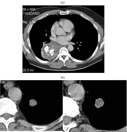
Figure 1.
Hamartoma. (a) Unusually large hamartoma with large chunks of ‘popcorn’ calcification. Initial diagnosis was made by biopsy and the mass was surgically excised. (b) A solitary nodule imaged on 5 mm (left) and 1.25 mm (right) slice thicknesses. The thinner section clearly demonstrates a significant amount of fat in this lesion indicating a hamartoma as the diagnosis. This could not be asserted with confidence on the 5 mm slice. The diagnosis was confirmed at surgery.

