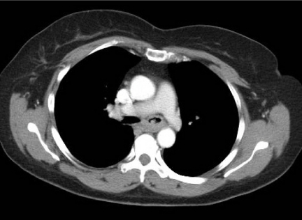
Figure 2.
Adenoid cystic carcinoma. Axial post-contrast CT image of a middle-aged female who presented with haemoptysis. There is abnormal soft tissue invading the left main bronchus and seen extending along the bronchus consistent with the submucosal spread often found in such tumours. Diagnosis was made at bronchoscopic biopsy.

