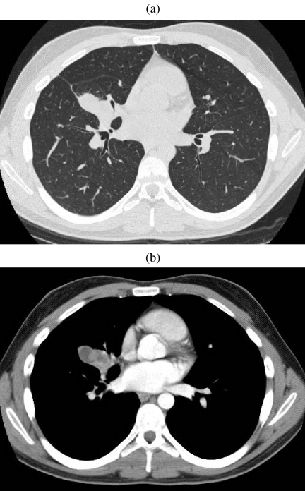
Figure 3.
Mucoepidermoid carcinoma. (a, b) CT on lung and soft tissue windows demonstrating a heterogeneous mass closely associated to the middle lobe bronchus. On bronchscopy, an expansile endobronchial tumour was seen. The diagnosis of mucoepidermoid carcinoma was established by bronchoscopic biopsy.

