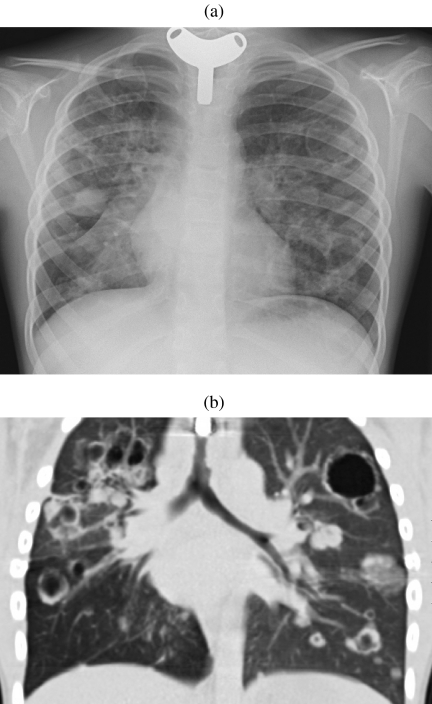
Figure 5.
Pulmonary papillomatosis. (a) Chest radiograph in a young adult with a previous tracheostomy for extensive laryngeal and pulmonary papillomatosis. Numerous nodules and thin-walled cysts are demonstrated bilaterally. (b) Coronally reconstructed CT of the same patient demonstrating tracheal plaques as well as numerous pulmonary nodules and cavitating masses. (Courtesy of Dr Catherine Owens, Great Ormond Street Hospital.)

