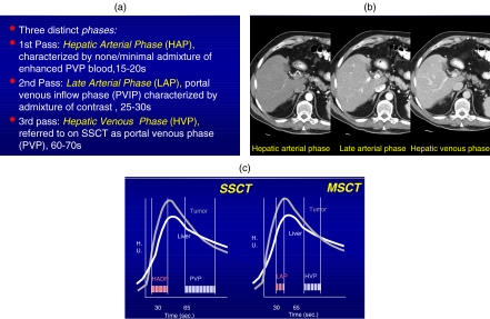
Figure 4.
(a) Definitions of the various contrast phases using MSCT. (b) Demonstration of contrast dynamics during the HAP, LAP, and HVP stages showing initially hepatic arterial enhancement while the second phase shows a mixture of hepatic artery and portal venous enhancement and the latest phase showing good portal venous enhancement. (c) The most effective terminology for MSCT is the late arterial phase (LAP) and the second phase, the hepatic venous phase (HVP) replacing the previous terminology (HADP and PVP).

