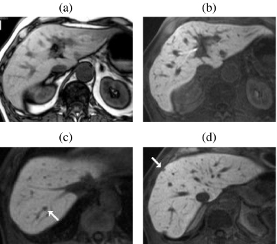
Figure 5.
Multiple metastases—improved detection with Gd-EOB-DTPA at the hepatocyte phase of enhancement. Compared with non-contrast T1w images (a) liver-to-lesion contrast is improved on 20 min post-contrast T1w images (b). Additional surgically confirmed sub-cm lesions (arrows) were only visible on hepatocyte phase images ((c) and (d)). Adapted with permission from Robinson PJA, Ward J (2006) MRI of the Liver: A Practical Guide. Informa Healthcare, New York.

