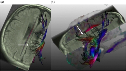
Figure 3.
Intracerebral arteriovenous malformation (AVM). (a) Tractography calculated from diffusion weighted imaging and registered on an axial FLAIR image shows the AVM with flow voids (arrow) and fibres of the corona radiata, including the cortico-spinal tract, which abuts on the AVM. (b) Sagittal view of the complete result of the tractography which superimposes the AVM (arrow).

