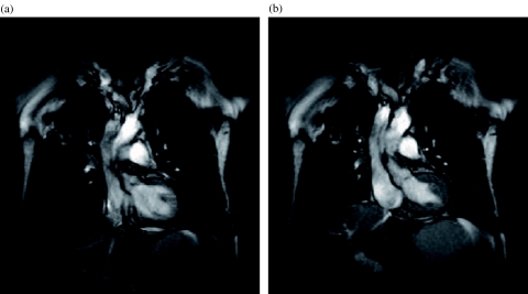
Figure 4.
Lung cancer. (a) Inspiratory image from a dynamic trueFISP sequence obtained with a temporal resolution of three images per second shows the tumour in the right upper lobe with a small clip artifact within the tumour. (b) Expiratory image from the same series shows the displacement of the tumour and also indicates the lack of chest wall infiltration.

