Figure 1.

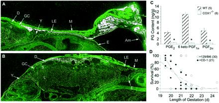
The etiology of prostaglandins in parturition. (A and B) In situ hybridization of 35S-labeled COX-1 (A) and COX-2 (B) in wild-type uteri on the morning of expected delivery (×10). Top includes implantation and interimplantation segments with the intact amniotic sac. Sections of the labyrinthine (L), spongiotrophoblast (S), and giant cell (GC), layers of the placenta and uterine luminal epithelium (LE) myometrium (M) and decidual (D) of the uterus are shown, along with fetal epidermis (E), amniotic membrane (Am), and yolk sac (Y). Sense probes were negative at sites of specific hybridization. (C) Prostaglandin concentrations of wild-type and COX-1−/− uteri on the morning of expected delivery, determined by GC mass spectrometry. *, P < 0.005. (D) Delayed parturition in 51 COX-1−/− litters on two genetic backgrounds. Mice with a 129/Bl6 background delivered later with less surviving pups, whereas CD-1 background mice had more severe lethality when parturition was delayed (129/Bl6 = dashed, R2 = 0.48; CD-1 = solid, R2 = 0.90). Overlapping points exist which represent the outcome of more than one litter.
