
Figure 3.
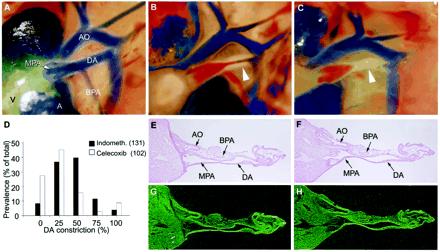
Prostaglandin effects on the fetal ductus arteriosus. (A–C) DA closure in untreated wild-type pups after delivery; immediate newborn period (A), 30 min–1 h (B), and 1–3 h of age (C) (×40). Arrowhead indicates closing DA. (D) Effect of COX inhibitors on in utero DA patency. Wild-type pups exposed to indomethacin or celecoxib before delivery were scored for the degree of DA constriction. (E–H) Brightfield (E and F) and darkfield (G and H) in situ hybridization images of 35S-labeled COX-1 (G) and COX-2 (H) mRNA in the fetal ductus arteriosus before delivery on the morning of day 19 (×40). Atria (A), ventricle (V), main pulmonary artery (MPA), branch pulmonary artery (BPA), ductus arteriosus (DA), and aorta (AO) are shown.