
Fig. 4.
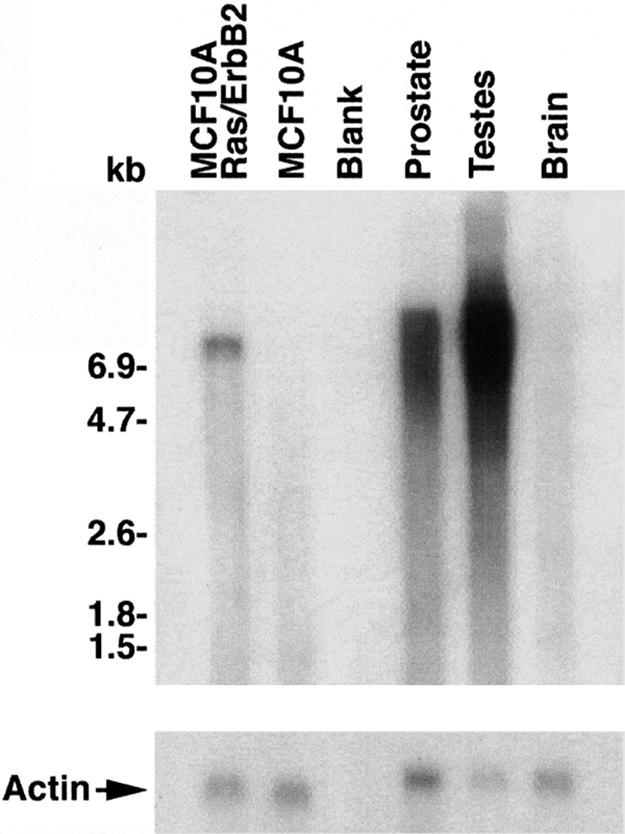
Analysis of POTE–actin fusion transcript in a breast cancer cell line. Northern blot analysis showing expression and transcript sizes of POTE–actin fusion transcript in different samples. Approximately 2.5 μg of mRNAs from different samples was run on agarose gel and transferred to a nylon membrane. The 1.2-kb probe was generated by PCR and labeled with 32P by using the random priming extension method. Membrane was incubated for 2 h in hybridization buffer followed by addition of denatured probe and incubation for an additional 12 h. Membrane then was washed and subjected to autoradiography. A specific band of ≈7.5 kb in size was detected in the MCF-10A-Ras/ErbB2 lane, but in prostate and in testis the signal is somewhat diffuse over 6.5 kb to 9.0 kb in size, and there are no detectable bands in MCF-10A and in brain.