
Fig. 5.
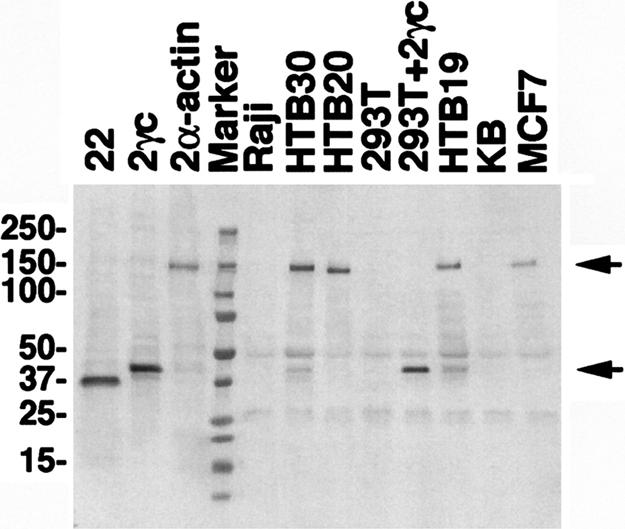
Detection of POTE–actin fusion protein in a breast cancer cell line. Cell lysates from different cell lines were subjected to IP by using HP8 antibodies. Immunoprecipitates were resolved in a 4–20% PAGE gel, transferred into a PVDF membrane and immunoblotted with PG5 antibody. A specific band of 140 kDa in size (upper arrow) was detected in MCF-7, HTB-19, HTB-20, HTB-30, and POTE-2α–actin-transfected 293T (positive control) lysates. No specific band was detected in KB, Raji, or 293T cell extracts. A smaller protein of 42 kDa in size (lower arrow) was detected in HTB-19 and in HTB-30, representing POTE protein without actin fusion. As positive controls for Western blot analysis, cell lysates prepared from 293T cells transfected with each POTE-encoding plasmid were loaded in the first three lanes without IP.