Fig. 2.
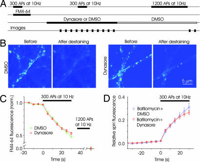
Dynasore has no immediate effect on exocytosis. (A) Schematic of the time course of FM-labeling experiments. A loading stimulus of 300 APs at 10 Hz was used to label synaptic vesicles with FM4-64. After the wash step, medium containing either 0.4% DMSO or 80 μM dynasore was added. After a 5–10-min wait, a destaining stimulus of 300 APs at 10 Hz was given, and images were obtained every 5 s. Normal solution containing DMSO was then perfused for 25 min to allow reversal of the effects of dynasore. A final round of 1,200 APs was delivered to release any remaining dye and to obtain baseline values of fluorescence. (B) Fluorescence image from sample experiments depicting FM-labeled boutons before and after destaining stimulus in the two conditions. (C) Time course of fluorescence change for synapses in DMSO and dynasore. The decay of fluorescence, an index of exocytosis, was similar for the two conditions in the early phase, indicting that dynasore had no immediate effect on exocytosis (n = 4 experiments, 200 boutons each). (D) Exocytosis measured with spH was unaffected by dynasore. Synapses expressing spH were stimulated (300 APs) in the presence of bafilomycin to get an index of net exocytosis. Fluorescence change was normalized to a total value obtained after neutralizing all vesicles with NH4Cl. Fluorescence changes in the presence of dynasore were similar to those in bafilomycin (dynasore: n = 3 experiments, 80 boutons; bafilomycin: n = 3 experiments, 70 boutons).

