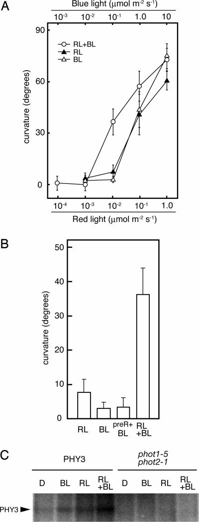
Fig. 3.
RL and BL signals are processed synergistically within PHY3. (A) Fluence rate response of hypocotyl phototropism. PHY3-expressing phot1–5 phot2–1 transgenic Arabidopsis (PHY3–1) was grown in the dark for 3 days, and then light irradiation was done as follows: RL, continuous unilateral RL for 16 h; BL, continuous unilateral BL for 16 h; and RL+BL, RL and BL were irradiated simultaneously for 16 h. At the end of the lateral-light irradiation, phototropic curvatures were measured. Each bar indicates a mean of 15 measurements with standard deviations. (B) RL enhancement effect on BL-induced phototropism. Phototropic response was induced as in A. Fluence rate irradiated was 0.01 μmol·m−2·s−1 of RL and 0.1 μmol·m−2·s−1 of BL. preR+BL, exposed to 10 μmol·m−2·s−1 of RL for 58 s before being given lateral BL. Each bar indicates a mean of 15 measurements with standard deviations. (C) Autoradiograph, showing the light-dependent phosphorylation of PHY3. Microsomal membranes prepared from dark-grown transgenic Arabidopsis expressing PHY3 (PHY3–1) or Arabidopsis mutant (phot1–5 phot2–1) were given a mock irradiation (D), irradiated with BL (at a total fluence of 2.1 × 10⁴ μmol·m−2), RL (at a total fluence of 1.1 × 10⁴ μmol·m−2), or irradiated both BL and RL simultaneously (RL+BL). All manipulations were carried out under dim green light.

