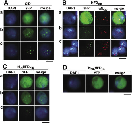
Figure 1.
Transiently expressed CID-YFP shows two coexisting patterns of localization, at centromeres and throughout chromatin. CID-YFP (A), HFDCID-YFP (B) and NH3HFDCID-YFP (C) can localize throughout chromatin (a), only at centromeres (c) and both, at centromeres and throughout chromatin (b). NCIDHFDH3-YFP (D) localizes only throughout chromatin. YFP is shown in green. DAPI-staining is shown in blue. The immunolocalization pattern obtained with αNCID, which specifically recognizes the N-terminal domain of CID, is shown in red only in (B). Bars correspond to 5 μm, except in row c of (B) where it corresponds to 10 μm.

