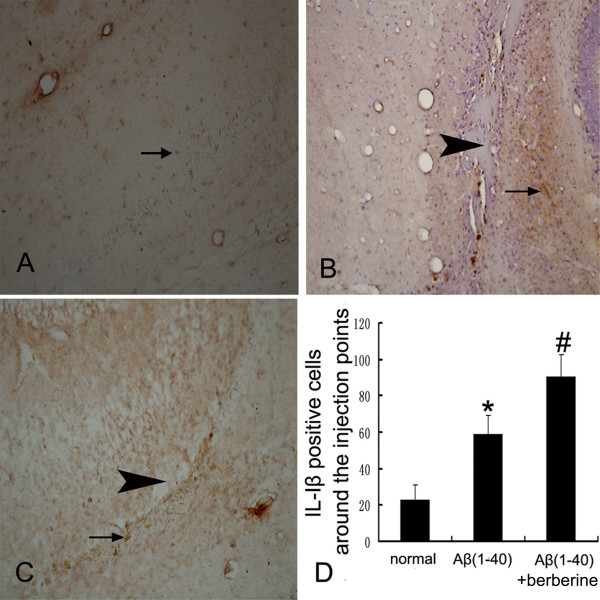
Figure 2.

Immunohistochemistrical evaluation of the effect of berberine chloride on the expression of IL-1β. (A) the expression of IL-1β(arrow) in the normal rat hippocampus.(B) the expression of IL-1β(arrow) around the injection point (arrowhead) in the Aβ(1–40) group. (C) the expression of IL-1β(arrow) around the injection point (arrowhead) in the Aβ(1–40)+berberine group.(D) the table represents the number of IL-1β positive cells around the injection point in 400× fields. Values represent the means ± SD of 6 rats.*P < 0.01 when compared with the normal group. # P < 0.01 when compared with the Aβ(1–40) group.(A,B,C: 200×)
