Figure 2.
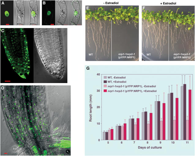
Subcellular Localization of YFP:NRP1 and YFP:NRP2 Proteins and Rescue of the Mutant Phenotype.
(A) and (B) Transgenic tobacco BY-2 cells expressing YFP:NRP1 and YFP:NRP2, respectively, were visualized by fluorescence confocal microscopy. YFP fluorescence image (left panels), bright-field differential interference contrast image (middle panels), and their merged image (right panels) are shown. Note that green fluorescence is concentrated in the spherical nucleus but absent from the nucleolus inside the nucleus. Bars = 10 μm.
(C) and (D) Root tip and stem-root junction region, respectively, from a transgenic Arabidopsis plant expressing YFP:NRP1. YFP fluorescence (in green) and differential interference contrast images are shown. Bar = 20 μm.
(E) and (F) Wild-type and transgenic double mutant nrp1-1 nrp2-1 (pYFP:NRP1) plants were grown in the in vitro culture medium in the absence or presence of the transgene expression inducer estradiol. Images were taken at 14 DAG.
(G) Comparison of root elongation between wild-type and the rescue-transgenic mutant nrp1-1 nrp2-1 (pYFP:NRP1) plants. The mean value from 20 plants is shown. Error bars represent standard deviations. Both YFP:NRP1 and YFP:NRP2 constructs were under the control of the induced estradiol-inducible promoter.

