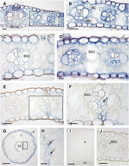
Figure 8.
In Situ Localization of Cytosolic GS Transcripts in Leaf and Root Sections of the Wild Type and the gln1-4, gln1-3, and gln1-3 1-4 Mutants.
(A) and (B) Localization of GS1 transcripts in the leaves of the wild type (A). Magnification of the zone containing the MC and BSC cells (B).
(C) Localization of GS1 transcrips in the gln1-4 mutant showing the absence of staining in the BSC.
(D) Localization of GS1 transcripts in the gnl1-3 mutant showing staining in the BSC.
(E) and (F) Localization of GS1 transcripts in the gln1-3 gln1-4 mutant.
(F) Magnification of the zone containing the BSC showing the presence of staining in the vascular tissue (arrow).
(G) and (H) Localization of GS1 transcripts in the roots of the wild type showing the presence of a strong signal in the epidermis and in the two outermost cortical cell layers.
(H) A magnification of the root central cylinder showing the presence of signal in the vascular tissue (arrows).
(I) Transverse section of a root tissue hybridized with the digoxygenin-labeled sense probe showing the absence of signal.
(J) Transverse section of a leaf tissue hybridized with the digoxygenin-labeled sense probe showing the absence of signal.
c, cortex; cc, central cylinder; e, epidermis. Bars = 100 μm.

