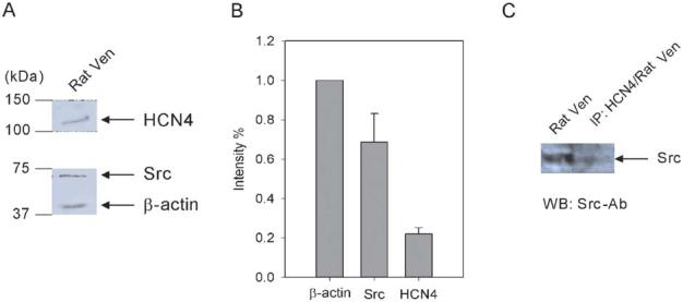
FIGURE 7.

Interaction of HCN4 with Src in rat ventricle. A, Western blots of HCN4 and Src proteins in rat ventricle. The membranes were cut in half. The two halves were incubated with anti-HCN4 and anti-Src antibodies, respectively. The half membrane fraction incubated with anti-Src antibody was washed and reprobed with anti-β-actin antibody to reveal the β-actin signal. B, Src and HCN4 expression levels are normalized to β-actin, which is used as an internal control (n = 3). C, Right lane: rat ventricular sample was immunoprecipitated by a specific HCN4 antibody followed by Western blotting using a specific Src antibody; left lane: Western blotting of rat ventricle sample using a specific Src antibody.
