
Figure 1.
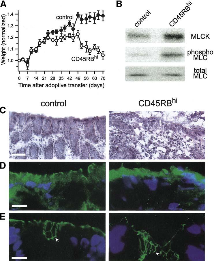
In vivo immune-mediated colitis is associated with increased epithelial MLCK expression, increased MLC phosphorylation, and tight junction disruption.
A. Adoptive transfer of CD4+CD45RBhi T cells (CD45RBhi) causes weight loss. Data are mean ± SE of four mice for each condition and are representative of 2 independent experiments. B. Colonocytes were isolated from control mice or after CD4+CD45RBhi adoptive transfer and analyzed by SDS-PAGE immunoblot for MLCK expression, MLC phosphorylation, and total MLC as a loading control. CD4+CD45RBhi adoptive transfer caused a 3.3±0.5-fold increase in MLCK expression and a 2.7±0.3-fold increase in MLC phosphorylation (p<0.03 for both). Data are mean ± SE of two samples from separate mice for each condition and are representative of 2 independent experiments. C.Histological examination shows that CD4+CD45RBhi adoptive transfer caused chronic inflammatory bowel disease with a moderate activity. Bar = 40 μm. D.Immunofluorescent analysis of occludin (green) localization in colon sections shows marked loss from the epithelial tight junction and appearance in intracellular vesicles after CD4+CD45RBhi adoptive transfer. Nuclei (blue) are shown for orientation. Bar = 10 μm. E. Immunofluorescent analysis of ZO-1 (green) localization in colon sections shows the development of an undulating network at the tight junction (compare areas marked by arrows) after CD4+CD45RBhi adoptive transfer. Nuclei (blue) are shown for orientation. Bar = 10 μm.