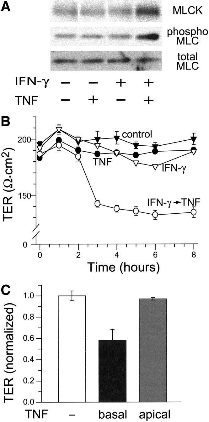
Figure 2.
IFN -γ primes Caco-2 monolayers for TNF-induced barrier dysfunction.
A. Caco-2 cells were pre-incubated with IFN-γ, 10 ng/ml, for 24 hours prior to transfer to fresh media, without IFN-γ, either with or without TNF, 2.5 ng/ml, as indicated. Monolayers were then harvested and analyzed by SDS-PAGE immunoblot for MLCK expression, MLC phosphorylation, and total MLC as a loading control. When IFN-γ-primed cells were treated with TNF, MLCK expression increased 2.1±0.1-fold and MLC phosphorylation increased 3.0±0.6-fold (p < 0.05 for both). Other treatments did not significantly alter MLCK expression or MLC phosphorylation. B. TER of control monolayers (black triangles) was measured for 8 hours after transfer to fresh media. Addition of TNF, 2.5 ng/ml, to the basal media had no effect on TER (black circles). Pre-incubation with IFN-γ, 10 ng/ml, for 24 hours prior to transfer to fresh media without IFN-γ, also had no effect on TER (white triangles). In contrast, the TER of monolayers pre-incubated with IFN-γ prior dropped within 3 hours after transfer to fresh media with TNF (white circles, p<0.01). Data are mean ± SE of triplicate monolayers. Data are representative of more than 10 independent experiments. C. All monolayers were pre-incubated with IFN-γ, 10 ng/ml, for 24 hours. Transfer to fresh media without cytokines (white bar) had no effect on TER, while transfer to media with 2.5 ng/ml TNF in the basal chamber induced marked TER loss (black bar, p<0.02). Transfer to media with 2.5 ng/ml TNF in the apical chamber had no effect on TER. Data are mean ± SE of triplicate monolayers 8 hours after transfer and were normalized to the overall mean TER of monolayers pre-incubated with IFN-γ. Data are representative of 4 independent experiments.

