Figure 3.
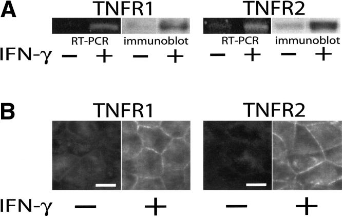
IFN-γ upregulates both TNFR1 and TNFR2 in Caco-2 monolayers.
A. RT-PCR and SDS-PAGE immunoblot analyses show that both TNFR1 and TNFR2 mRNA and protein expression are upregulated after 24 hours of culture with IFN-γ added to the basal chamber (p<0.01 for both). Data are representative of 4 independent experiments, each with duplicate samples. B. Immunofluorescence of monolayers before or after 24 hours of culture with IFN-γ confirms increased surface expression of TNFR1 and TNFR2. The images shown were taken at focal planes basal to the tight junction and TNF receptors were only detected on lateral membranes. Exposures and post-processing were identical for images of each TNF receptor. Bar = 10 μm.

