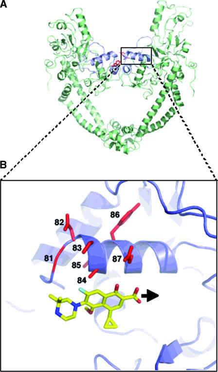
FIG. 2.
(A) Ribbon representation of the 59-kDa N terminal of GyrA adapted from the crystal structure of the breakage-reunion domain of the GyrA protein of E. coli (16). The QRDR (quinolone resistance-determining region) is in blue, and the active site (Tyr122) is in red. (B) Close-up of the region outlined by the broken box highlighting the schematic representation of the fluoroquinolone positioning in the GyrA α4 helix domain. The fluoroquinolone used for the model is gatifloxacin. The black arrow shows the displacement of the fluoroquinolone positioning in the M. tuberculosis GyrA α4 helix domain compared to the one proposed for E. coli as suggested by our study.

