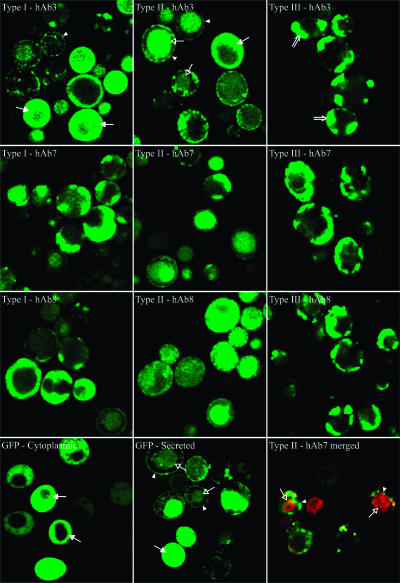
FIG. 9.
Representative confocal images of cells expressing GFP and fusion proteins. The cells were induced for 3 days prior to microscopy. GFP was expressed in both cytoplasmic and secretion systems for comparison with intracellular distributions of fluorescent fusion proteins. For type II hAb7, total protein was labeled with an anti-c-myc antibody, followed by anti-mouse Alexa Fluor 555 (red), and GFP fluorescence (green) was used to monitor the localization of fluorescent fusion proteins. Alexa Fluor 555 (red) and GFP (green) fluorescences were merged to show colocalization. Typical patterns of GFP fluorescence are indicated as follows: arrowheads, small surface punctate; open arrows, vacuolar localization; solid arrows, cytoplasmic localization; double-line arrows, large surface punctate.

