Figure 1.

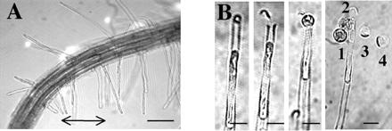
Recovery of plasma membrane at the apex and subapical region of young root hairs. (A) Apex of a 3-day-old A. thaliana root. The arrow marks young, growing hairs used for experiments. (Bar = 100 μm.) (B) In situ laser microsurgery and recovery of hair spheroplasts (12, 13). (From left to right) Root hair tip plasmolysis; tip cell wall cut by laser; recovery of apical plasma membrane by hair deplasmolysis; recovery of subapical plasma membrane by stronger deplasmolysis. Numbers mark the order of spheroplast extrusion from the tip. (Bar = 15 μm.)
