
Figure 2.
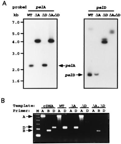
(A) Genomic Southern blots showing the absence of native genes and appearance of new fragments from the disrupted genes in the mutants. Genomic DNA (10 μg each) was digested with HindIII and probed with cDNAs of pelA (Left) and pelD (Right). Size markers shown on the left apply for both panels. (B) RT-PCR showing the absence of transcripts from the disrupted genes and the presence of transcripts from the native genes. Gene-specific RT-PCR amplification of pelA, pelB, or pelD was done by using gene-specific primers (20) and RNA isolated from N. hematococca-infected pea epicotyl. WT, wild type; ΔA, pelA disruptant; ΔD, pelD disruptant; and ΔA, ΔD: pelA and pelD double-gene disruptant. Position of the expected products from the three genes are shown by the arrows. Molecular size marker (M), 123-bp DNA ladder.