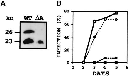
Figure 3.

(A) Immunoblot analysis of proteins from N. hematococca wild type (WT) or pelA disruptant (ΔA) grown on pea epicotyl sections showing products of pelA (26 kDa) and pelD (23 kDa) in WT and only pelD product in the pelA disruptant. After 5 days of growth at 24°C (28), the soluble proteins were subjected to immunoblot by using anti-PLC (29) and enhanced chemiluminescence detection system. (B) Time course of infection of pea epicotyl sections by WT or pelA disruptant in the presence of antibody to PLA or preimmune IgG. Assay was done with 104 conidia and 10 μg of IgG (28, 31). Infection in the presence of water only was identical to that in the presence of preimmune IgG. (○) WT, preimmune; (□) ΔA, preimmune; (●) WT, anti-PLA; and (■) ΔA, anti-PLA. % infection, % of epicotyl sections that showed lesion.
