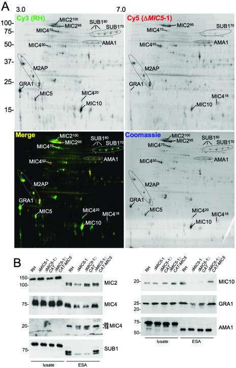
FIG. 2.
Loss of MIC5 expression changes the ESA secretory profile. (A) Large-scale ESA fractions were isolated from ΔMIC5-1 and RH parasites, normalized as to protein content, and labeled with either cy5 (red) dye or cy3 (green) dye, respectively, before protein populations were combined and run together on 2-D SDS-PAGE. Following laser-scanning acquisition of the fluorescent images, the gel was stained with Coomassie blue dye to label all proteins. Top panels are images taken in single channels, whereas the bottom left panel is merged and the bottom right is the Coomassie-stained gel. Positions of various secreted proteins are indicated based on direct identification by mass spectroscopy or their migration relative to the previously described map of the Toxoplasma ESA proteome (44). (B) Western blots of parasite lysates and ESA fractions from RH, ΔMIC5-1, ΔMIC5-1::CAT, and ΔMIC5-1::CAT-MIC5 strains probed with anti-SUB1, anti-MIC2, anti-MIC4, anti-MIC10, anti-GRA1, or anti-AMA1 antibodies.

