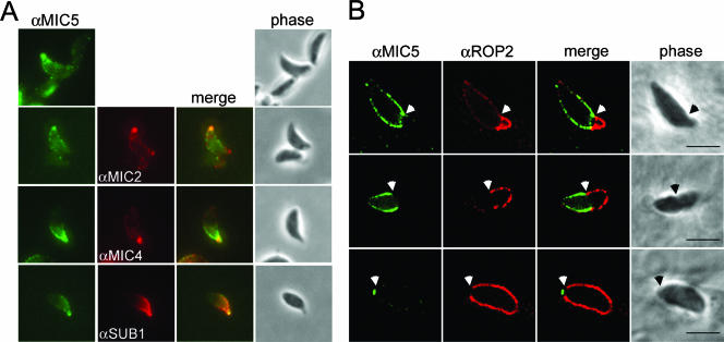
FIG. 4.
MIC5 is exuded onto the parasite surface and labels a region near the moving junction during host cell invasion. (A) MIC5 partially colocalizes with MIC2, MIC4, and SUB1 on the surface of extracellular parasites stimulated to secrete micronemal contents. Extracellular parasites were stimulated to release micronemal contents, fixed, and subsequently labeled with anti-MIC5 (αMIC5), anti-MIC2 (αMIC2), and anti-SUB1 (αSUB1). (B) MIC5 labels a region near the moving junction between the parasite and host cell plasma membranes during invasion, treadmilling toward the posterior of the parasite as it penetrates into the PV. RH parasites added to HFF monolayers in a pulse were trapped in a partially invaded state and semipermeabilized to examine the contents of the PV without interference from signal within the parasite. Anti-ROP2 (αROP2) antibodies denote the PV. White arrows indicate the position of the moving junction. Bars, 5 μm.

